Free On-Demand Webinar
with Dr. Robert Shiroff
Renowned Cardiologist and CompuMed Chief Medical Director, Dr. Robert Shiroff presents a webinar on the unique role of Heart Catheterization in Donor Management. This is both practical and technical, touching on the why and how of improving donor management through heart catheterization.
Topics include:
- Importance of heart cath in overall donor management
- Donor focused interpretation of the procedure for potential heart placement
- Reading and review of heart cath
- Insights on how to increase the number of transplantable hearts
Who is this for?
Donor and Transplant Professionals
Key Takeaways:
- Minimum two views of the Right Coronary Artery, Left Anterior Oblique (LAO) and Right Anterior Oblique (RAO).
- Multiple views of the Left Coronary Artery with adequate unlapping of major branch points (left anterior descending and left circumflex arteries).
- Documented pressure measurements, oxygen saturation and cardiac output for Right Heart Catheterization.
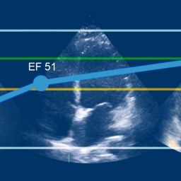
- High quality RAO projection cineangiogram of the left ventricle (adequate for estimated EF%).
Dr. Robert “Bob” Shiroff
CompuMed Chief Medical Director
CompuMed is honored to have Dr. Shiroff on our team as he serves the donor and transplant community. His in-depth knowledge and expertise helps assess, manage, and improve the recovery of donor hearts, resulting in an increase in the number transplanted into needy recipients. Knowing that each recovered heart has the opportunity to save a life is humbling and extremely rewarding. We are grateful for his passion to share his knowledge and experience with the OPO and Transplant community.
Interested in other topics? Let us know!
[00:00:00] Laura Carroll: Hi, everyone. I believe most people have signed on. Hello. I am Laura Carroll, and on behalf of the entire CompuMed team, thank you for joining us for another, in our expert webinar series. So for many of you, Dr. Robert Shiroff doesn’t need an introduction. You may have spoken with him at 2:00 AM, multiple times, and many of you just might have his cell phone number in your contacts.
[00:00:26] Dr. Shiroff is a renowned cardiologist with so much history and experience, we could actually have a webinar just on his content alone. He’s been part of the CompuMed team since 2013, reading echoes for OPOs, leading development of protocols for these reads, helping customers, running our QA, and so much more. By our estimate, Dr. Shiroff has read more donor echoes than any other doctor ever. And with all of his experience, every time we work with him, we learn something new. While we were preparing for the heart cath, we actually learned that as a senior fellow, he was teaching first year fellows, heart cath procedures. And we also hear that his supervising doctors actually allowed him to perform caths early on without supervision.
[00:01:13] Dr. Shiroff’s passion for helping all of you save more lives is endless. And to quote him, the saving of a heart is a saving of a life. Without further introduction, Dr. Shiroff.
[00:01:25] Dr. Robert Shiroff: Thanks, Laura. It’s a pleasure to be here and thanks the kind introduction. Today’s topic is heart cath and donor management, and I thought I’d begin with a little history and start with the discussion about William Harvey in 1628, who described the circulation of the heart.
[00:01:43] There’s a picture here. The heart circulation. It was described by Dr. Harvey in 1628. And if you begin with the blood flow and the left ventricle, you see here, this, the pumping chamber where all action begins and the one that we’re most concerned about in terms of function when we look for heart transplant. Blood comes out of the left ventricle through the aortic valve, the first vessels coming off the aorta out of the aortic valve are the left coronary artery and the right coronary artery. The blood then circulates into the aorta and goes to the brain, the upper body and the lower body, the kidneys, the liver in every organ in the body.
[00:02:17] Blood then comes back after going through smaller and smaller arteries, and organs, in the form of capillaries. One cell thick tubes and oxygen and food is then exchanged for the waste products and CO2. Blood is then returned through small veins to larger veins. And up from the bottom of the, distal lower extremity through the inferior vena cava from the upper extremity through this, through the superior vena cava into the right atrium.
[00:02:43] The blood then goes from the right atrium across the tricuspid valve into the right ventricle, across the pulmonic valve where it’s pumped out into the lungs through the pulmonary arteries. The pulmonary, the lungs then exchange oxygen and CO2 at the capillary level. And the blood is then returned through venules into the pulmonary veins, which dump its blood into the left atrium, and it crosses the mitral valve into the left integral and the cycle continues. So it’s a nice circuit where blood goes back and forth. I review this and many of you probably know this, having gone through nursing school, but I thought it’d be worthwhile just to review for purposes of, bringing everybody to the same level.
[00:03:21] History of cardiac catheterization begins with the right heart cath by a guy named Wegner Forsman. Dr. Forsman was a 25 year old, newly minted doctor in Germany and he decided that he wanted to prove that you could reach the heart from the peripheral circulation of the vein.
[00:03:37] He had a young lady he was friends with and he convinced her to help him. She was kind of like a radiology tech at the time. And radiology was fairly new in its infancy at that point, taking pictures. That’s Dr. Forsman here on the left. He went to the lab and he told her he would do it on himself.
[00:03:54] And she said, no, no, no. She loved him. Didn’t want him to die. And she wanted him to do it on her. So he faked doing on her. He numbed her arm up and made a small incision on her arm and faked it, and then did his own arm and put the catheter up. He then got her back where she should be and they went down a floor to the radiology suite and he took a picture of this catheter.
[00:04:14] That actually is a urinary catheter that he put in from his antecubital fossa that went up through the axillary vein in the subclavian vein, into the superior vena cava, into the right atrium. And here’s the tip of the catheter sitting. It’s the first example of right heart catheterization being done in 1929.
[00:04:32] Following that in 1940s, left heart catheterization was proven to be successful by Drs. Cournand and Richard. And, then in late 1960s, a guy named Mason Sones, and I was privileged to meet Dr. Sones at a meeting and going out to dinner with him and had a few drinks.
[00:04:49] And he proceeded to tell us how he discovered the success and was successful in doing cardiac catheterization. He over a few drinks said that he was doing a left heart catheter. And at that time they put a catheter that had end holes and a side hole. Now a catheter is a plastic tube that is hollowed out and he put a catheter with that which had end hole, the end of the catheter, so he could measure pressures, and the side hole so if you injected dye, it wouldn’t come out as a jet; it would come out of all the three or four holes that were there. He, we were doing this for several years from, the forties, on. He put the catheter in. He was sure he was in the right spot. Didn’t do an injection, got ready to do the injection and he did a power injection through a power device at that time. And he did it. And lo and behold, he was in the right coronary, a huge right coronary artery and the patient didn’t die. He was so excited about this, that he decided that if he could do it accidentally and not kill the patient, he could do it on purpose.
[00:05:43] And that was the initiation of selective coronary arteriography, which led to aortic coronary bypass graft surgery, first being done at Cleveland clinic in the early sixties, with the help of bypass machine that was invented at Jefferson by Dr. Gibson. So the history of that, we’re talking about dates back for many years now. And, I was fortunate enough to have met, one of the guys who started the whole thing, was quite an experience. He was quite the character.
[00:06:08] The indications for cardiac catheterization and a patient who’s a potential transplanted heart, pretty much everyone agrees, over 40 year old should have a selective cardio arteriography. History of unknown heart abnormalities will precipitate the investigators wanting to know, the transplant centers wanting to know what you’re telling us something, we don’t understand it. The echo, we can’t tell if there’s an abnormality there, so we want to see a heart cath. We want to see the coronaries. The major one, besides over 40, is substance abuse with methamphetamine, cocaine, which can affect coronary blood flow and affect the coronary arteries, per se, and also cause myocardial injury.
[00:06:43] Other things that are coming into Vogue now, and I think are gonna be more so as we get further, along in this adventure is the significant risks for heart disease. We’re seeing younger and younger diabetics in the United States with the obesity epidemic that’s present in the United States.
[00:06:59] And because of that, If you have hypertension, diabetes, hyperlipidemia, and you have a bad family history, the chances of having coronary disease are higher. And the reason the transplant centers are very concerned about this is that the progression of coronary disease is much more rapid. Once you transplant a heart it’s related to rejection issues, as well as the medications used for rejection treatment.
[00:07:23] I once had a family of hyperlipidemic patients who had all of them had bypass surgery in their twenties. So you can tell it isn’t just a disease of older people, and it’s gonna becoming a disease of younger and younger people as the diabetes and hypertension epidemics continue in the United States.
[00:07:39] This is a right heart catheter here. It was designed by two doctors in Cedar Sinai, Dr. Ganz and Dr. Swan. I’ve had the privilege of working with Dr. Swan on a couple of legal cases, and it’s a fascinating catheter. They decided that it was really neat if you put a balloon on the end of a catheter and could get it into a big enough vessel, it would float, the way the blood flows and because there’s this movement of blood in a single direction in the vein. And he, they put, they decided that was a clever idea. They also decided that it was a clever idea to measure temperature differences from one place. If you injected in the right heart, in the right atrium and you then sense the temperature difference in the pulmonary artery, you could then determine by complex calculations, cardiac output.
[00:08:23] So this catheter has multiple ports. One is the proximal port where the atrial dumps out into the atrium. It has a, distal port here, and this is central another port, which is available on some of the catheters. It has thermistor measurements here, and a plug into the electrical system here. So you at least have four or five ports minimum when you have these catheters.
[00:08:46] And it depends on what needs you have. So a right heart catheterization is done very simply by putting the catheter in and let me show you a right heart catheter. This is a catheter there. The catheter at this point is coming up from the inferior of vena cava into the right atrium. And it crosses the tricuspid valve into the pulmonary artery, into the right ventricle.
[00:09:08] You can see the balloon here and what’s gonna happen as I run this a little further is you’ll see the balloon pop up in the main pulmonary artery, because that’s how the blood goes. The right ventricle, squeezes, the blood and it pops it across. As I further advance this, the catheter then goes out further into the pulmonary artery and into what’s called a wedge position.
[00:09:27] The pulmonary capillary wedge pressure is critically important for knowing what left heart pressures are. The left end particular end diastolic pressure estimate is best done through a pulmonary capillary wedge pressure other than direct measurement. So on the way out of this whole thing, when you let the balloon come back, you deflate the balloon, the catheter withdraws a little bit, because it’s being pushed out there by the balloon. You measure pressures in the pulmonary artery. You get an oxygen saturation because you want to know what the sat is. So you make sure you have no shunts in the heart, like at ASD or patent foramen ovale. You then pull the catheter in the right ventricle, measure pressures there into the right atrium measure pressures there and get saturations.
[00:10:06] If you can, in the superior vena cava and inferior vena cava, if you can’t at least in the mid right ATR. And at that time, usually a catheter is in the aorta, because you’re gonna do coronaries and you get a saturation done from and a pressure done from that measurement as well. And that’s a complete heart catheterization.
[00:10:23] Left heart catheterization is, putting a catheter into the left ventricle. The catheters that are typically used, and have been for years, is called a pigtail catheter. It looks like a pigtail. This is the catheter here. This is one that I had an example of from when I was doing these procedures a bunch of years ago, and I stole a packet for teaching my son’s high school and college classes. I did physiology lectures for them, for the teachers. The other two catheters are preformed left coronary catheters. This one is the right coronary catheter, and this is the left coronary catheter, preformed catheters.
[00:10:57] It’s because of the shape of the aorta and how the arteries come off of the aorta, these catheters make it easy for the cardiologist to selectively catheterize the main arteries and get pictures of them. So what’s done here is this catheter is advanced. The pigtail catheter is with a wire in it, initially. You pull the wire out as it gets into the central aorta and you push it against the aortic valve. And when the valve opens up to allow blood to come out from the left ventricle, the catheter pops across the aortic valve with no untoward events occur usually, and it sits in the left ventricle.
[00:11:27] You then put dye through the catheter. Radiopaque dye, which then you do an angiogram and get a picture of the left ventricular chamber. And from that you can get a good estimate of left ventricular ejection fraction. You can also get objective measurements where if you have the proper computer equipment. Again, at the time of the cath drawing is made and you can get an objective ejection fraction.
[00:11:49] Most cardiologists rely on their eyeballs at this time because they have confirmation from echo as well. But this is the gold standard still for left ventricular function measurements. I’ll show you a couple pictures of an angiogram. This is a typical angiogram. These pictures, by the way, are all from OPO echos, OPO angiograms, excuse me. This is a, lousy angiogram, cuz you can hardly see the heart. And it’s a loop, so you can see that you can get an estimate of what’s going on, which it’s a best guess because you can’t see the margins because there’s not enough dye given. This is a cardiologist doing a hand injection with a syringe, trying to get opacification of the LV.
[00:12:24] He did it twice more. The second one was just like the first one. I didn’t bother showing that one. Here’s the third one where he gets a little better and you can actually see the chamber there. The chamber is right here coming around here. And it’s a better example, but it’d been better if he’d used the power injector and spent the extra two minutes to inject 15 CCS as a bolus. And then we would’ve gotten one good beat or two good beats and been able to see the heart much better.
[00:12:50] Selective coronary arteriography is the gold standard for looking at coronary anatomy. And here’s a picture of a coronary anatomy from a graphic point of view. The right coronary artery comes off of the right coronary cusp and runs down and gives a rise to a posterior sending coronary artery, typically. That you may see in reports that you’ll see that I dictate or my associate’s dictate or other cardiologists that there’s dominance mentioned. Right coronary dominance means that the right coronary posterior descending artery comes off of the right coronary artery. That happens in 70, 80% of the time in 5 to 10%, you have a left coronary dominance, which means it comes off the left circumflex, the posterior descending, and 10 to 20% you have co-dominant where you have two vessels, one from the right and one from the left circumflex, typically, that feed that same distribution. Why is that important? Well, it’s important because a branch that comes off of that artery supplies, the av node. It’s called the av nodal artery which, has a lot to do with electrical activity of the heart. And that’s why dominance is mentioned all the time in reports.
[00:13:52] The other artery comes off a left main, off of the left. Aorta is the left main coronary artery, which comes off a left coronary cusp. It gives rises to a left anterior descending, coronary artery, and multiple diagonal branches and a left circumflex that usually goes posteriorly and gives rise to obtuse, marginal branches in the posterior and lateral wall.
[00:14:11] So the whole heart is covered. In addition, small atrial branches a lot of times come off of the radio coronary artery and the left circumflex that supply the atrial walls. Since the major portion of the blood supply, the heart is and goes to left ventricle because it’s the thickest and requires the most blood supply.
[00:14:28] Next, you’re gonna see a whole bunch of coronary angiograms here. The arteries are tubes. They are three dimensional structures. If you think of a straw, if you hold a straw up against the wall and you take a picture of it, you can’t tell how thick the straw is, because it’s only a two dimensional picture in the three dimensional world.
[00:14:47] So what we do to compensate for that is we move the camera around the patient and take multiple views of that same artery. This is the left anterior descending coronary artery coming down here. The left circumflex is coming off and it actually is going posteriorly. This vessel over here is actually behind the heart and you’re seeing it as if it was in the same plane, because we’re looking at two dimensions.
[00:15:09] I’ll show you a couple more pictures and be able to see better that the left circumflex on this view is now coming off the back of the heart. You can see here this is the posterior wall and here the left circumflex is coming back here, and the left anterior is descending is over. Here’s the LAD now, it’s a much different view and there’s a diagonal branch coming off the left anterior descending right here. This is a normal from a, again, from a, an OPO angiogram. This is a right coronary artery. And again, many times when we get angiograms performed in the middle of the night by a cardiologist who’s not an eager player in the game that they do a single view of the right coronary and say, it’s normal.
[00:15:50] That’s really against the rules as far as I’m concerned, because again, it’s a two, three dimension. You can’t see in two dimension. It’s the same as holding a stack of paper up and against the wall. I’m looking at it. You cannot tell how thick the stack of paper is. It could be one piece thick, or it could be six inches or six feet thick.
[00:16:06] You can’t tell until you move around and look at your eye to see the thickness of the wall thickness of the paper. So here’s a right coronary artery. This is a dominant right coronary gives rise to a posterior descending coronary artery and a posterior lateral brain branch here. Again, these are normal.
[00:16:25] Here’s the two pictures of the right and see how different they look in different views. And you can tell their different views by where the spine is. Here’s the spine. It’s almost an AP view here. This is more of an RA, an LAO view. You can see these RV marginal branches are much better seen, and you can see the branch points.
[00:16:43] If I stop it, I’ll show you here. You can see branch points if you move it a little bit. Here you cannot see branch points. You cannot tell whether it’s normal coming off of that artery or not. And there’s one of those atrial branches I was talking about here, coming off, up here in the, in this, view.
[00:16:59] So at least two views of the right coronary artery should be accomplished in every study. Here’s a picture with some disease in it. We don’t always get, not everybody’s normal. The reason you do this procedure is because you wanna see whether you have disease or not. This is a 50- year- old gentleman who was potential transplant patient who had a good heart.
[00:17:16] Good ventricle. Good ejection fraction by echo, normal valvular structures by echo. No significant wall thickness abnormalities. Here you can see what looks like a narrowing to my eyeball, comparing this, the thickness of the wall here versus the thickness of it here, the diameter of it, and up here is of concern to me.
[00:17:34] I can’t tell about up here, although it’s a little bit more concerning. This is to me, this looks a little broader than this does. So they move the camera around, take another picture. And here it doesn’t take a rocket scientist to see that you’ve got a significant narrowing of that portion of the artery.
[00:17:51] You can see it much easier here, and here now you see there’s getting more significant narrowing in that or that part of the left anterior descending. This is the left circumflex. And to show you how different things are, a third view, which is called an axial view, where you shorten the, you shoot the picture from the feet to toward the head. You angle the camera as well as, a rotational, it, moves anterior posteriorly, from the feet toward the head. This is that area of narrowing that was so far down the artery. Now you can just about see it, but it’s way up north because of the angulation. And here, they’re trying to look more at this anterior descending and it was a good angiographer that defined that narrowing, and it’s a significant narrowing as well. So this heart was not a transplantable heart.
[00:18:36] Here’s the one that’s fun because, why would I show a left coronary and a right corner at the same time? The reason I show it is because if you look right here, there’s a haziness to the area here. And if you look here before it injects that haziness is still there. That’s calcium in a coronary artery.
[00:18:54] It’s a marker of disease. That disease, that artery doesn’t look too bad. It had some disease in it, but the calcium would probably make it not a transplantable heart. As it suggests that there’s the real disease there.
[00:19:07] See the calcium and it fills up with dye looks how different it looks. And you can see a narrowed artery here, narrow a little bit narrowing right there, and some tapering in here as well.
[00:19:26] Well, what kinda reports are you in? Can you expect to get from the cath lab? Every, every hospital’s a little different. This is one of the hospitals in the OPO referral business. And they put out a picture of showing the pressures in the chambers, and the saturations and the written words would be the, cardiac output and the oxygen saturations.
[00:19:47] And then multiple pages of descriptive of what happened in the cath lab. Every T is dotted, every I is dotted. Every T is crossed, showing every with every injection, what time it occurred, how many CCS were used, how much extra exposure there was, et cetera, et cetera, et cetera. And these are as long as 15 to 25 pages and they’re of little value except the pressure and the numerical numbers. The numerical pressure measurements and the cardiac output and saturations are the ones in the peripheral, vascular resistance and systemic vascular resistance are the numbers we want to have. So if you’re gonna send, if you want me to help or me and my associates and when you want us to look at this catheterization report for a right heart cath and left heart cath, please try and provide us with this information.
[00:20:33] The other thing you’ll get is many times is a report from the cardiologist, a preliminary report, he’s dictated it, which may take a long time to become typed. And that’s part of the reason I think we get to reinterpret these things, but this is a preliminary report done by a cardiologist, with the pressures, with the left ventricular pressures and the o2 sats and the cardiac outputs with the fact that he thinks it’s a normal coronary arteries, no CAD, and a generic picture of a coronary anatomy, which doesn’t help the surgeon to know if there’s any anomalies or not. But the, assumption would be that this is a normal coronary anatomy. Doesn’t say whether it’s right dominant or left dominant which is important for the transplant surgeon to know as well.
[00:21:16] So this isn’t optimal, but it’s acceptable. And if you get a full report, the transplant centers prefer to have a complete report, like the one that we dictate which describes the anatomy and, the pressures, and then summarizes it in the conclusions.
[00:21:34] I’ll stop here and let you ask some questions and then we’ll go on from there.
[00:21:39] I wanted, let me, one more thing I wanna make mention of is to take key takeaways and that is minimum of two views of the right coronary artery and the right anterior oblique and left anterior oblique projections. Multiple views, the left coronary artery with adequate unlapping of the major branches, the LAD with its diagonal branch and septal perforators should be unlapped, the circumflex with its obtuse marginal branches should be unlapped. And you document right heart catheterization with a optimally documented with right heart pressures, where you get PA pressures, RV, RA, SATs from the PA and, right atrium minimum as well as the aorta and cardiac output.
[00:22:20] For the cineangiography of the left ventricle, you want an RAO projection, usually in 30 degree angle and to the, with adequate opacification for estimated EF by the cardiologist, who’s reading it. If you get that, this, these four things on a cardiac cath, any qualified cardiologist, who does an intervention or invasive cardiology can interpret these things and come up with a report.
[00:22:46] The key then is to get the report as soon as possible so we can move forward with the transplant because we know all know the time is critical in these issues. I’ll stop here and let’s answer some questions and see how we do.
[00:22:58] Webinar Host: Thank you, Dr. Shiroff. Now we’re moving into our Q&A, so if you haven’t submitted your question yet, the Q&A section of Zoom can fill up fast.
[00:23:06] Some of you sensors in early, so Dr. Shiroff will be taking those first. So here we go. Dr. Shiroff, what is the minimum data you would like to see from a cath lab report?
[00:23:17] Dr. Robert Shiroff: Well, I just went over it pretty, fairly, I think. I could do it again. We can go back one slide and just make, mention this again, because I think it’s that important.
[00:23:25] The minimum from the heart cath and the right heart cath start with the right is pressures from the left of it. The pulmonary artery and mean pressure wedge pressure, right ventricular pressure, right atrial mean pressure, and oxygen saturation done in both the pulmonary artery main and in minimum main, the mid, right atrium. Along with an o2 sat from the aorta, central aorta, ideally. With those 3 o2 SATs, you can pretty much exclude a shunt lesion from the, in the right side of the heart of the HASD or VSD. Next thing you wanna make sure you get is a good left ventricular injection. If you’re gonna do an LV, if you’re gonna be there, you might as well shoot a picture of the left ventricle to see what the chamber looks like. And the adequate opacification is critical. A hand injection can be, is sometimes be very problematic and getting good pictures and being able to estimate the ejection fraction. The third thing, and third and fourth things on here is two items of the coronary anatomy. The right coronary artery, you should shoot in at least two pictures.
[00:24:27] If there’s any question about branch point of the posterior descending or posterior lateral branches of the or, the main right coronary artery, where it bends, one should shoot more pictures, because it’s critically important that we get a good look at these arteries. And the left coronary artery, the left main where it bifurcates into the left anterior descending and left circumflex and the anterior descending in its branches that major diagonal branches and left circumflex in its major obtuse marginal branches should be well opacified and the branch point should be well seen. That’s the minimum from a hard cath, as far as I’m concerned for adequate interpretation.
[00:25:02] Webinar Host: Thank you. Now, are there other options available for defining coronary anatomy besides selective coronary arteriography?
[00:25:10] Dr. Robert Shiroff: Yes, there is.
[00:25:11] Laura Carroll: Uh,
[00:25:11] Dr. Robert Shiroff: CT and geography is available.
[00:25:14] It is done with a 64 slice CT machine. One can get pictures of coronary arteries. They are not quite as good as, or accepted at this point in time in the literature; it’s, coming around. It requires special skill to interpret them and special training. You have to have great manipulative skills with the computers in order to interpret them, because you have to move the pictures around like crazy to get adequate opacity or adequate views of them. But yes, you can determine whether there was coronary disease. That, with a CT calcium score would be almost comparable to an angiogram. CTA can be done with a high powered CT machine, to answer your question.
[00:25:56] Webinar Host: Thank you, Dr. Shiroff. Genevieve Springer, founder and CEO at Oregon -AI asked, “To increase the number of transplanted hearts and other organs, how helpful would an instant precise whole organ tissue assessment be for the process at recovery and post transit?”
[00:26:16] Dr. Robert Shiroff: Wait. I’m not sure I’m qualified to answer that. I think I would defer to a lot of other experts besides myself to answer that question and I’m not sure what, she means. Repeat the question with this tissue.
[00:26:28] Webinar Host: Sure. “To increase the number of transplanted hearts and other organs, how helpful would an instant precise, whole organ tissue assessment be for the process at recovery and post transit?”
[00:26:41] Dr. Robert Shiroff: A whole organ tissue evaluation, I’m not sure what that means. I’m not privy to that information and, I’d have to defer that one. I can look it up and see if I can come up with an answer for her and ask some of my good friends here to help me out. But I, don’t know the answer to that. I think I, I don’t know is the answer.
[00:27:00] Webinar Host: Okay. Our next question comes from Susan Nelson, Director at Oregon Clinical Operations at Life Gift. She said, “Thoughts on use of beta blockers and heart donors?”
[00:27:12] Dr. Robert Shiroff: Well, we recently had an interesting echo that was done on a patient who was tachycardic and hypertensive at the beginning of the echo, and the left ventricular function was normal by the echo technician’s study. In the middle of the study, the patient received the IV metoprolol, IV beta blocker, and all of a sudden the ejection fraction dropped into the 30%, and the echo technologist recognized that it had happened and waited a few minutes and repeated this study part of the study again, and the ejection fracture was back up to normal. Beta blockers are certainly indicated and useful in multiple settings in multiple clinical settings and in, and should be customized to choose. I don’t think as a general rule, everybody should get a beta blocker. If you have tachycardia, it’s not controlled and you need to slow them down. Because it’s a significantly abnormal tachycardia. If a blood pressure is not controlled with other standard drugs, beta blockers can be helpful. For rhythm disturbances, beta blockers can be very useful, but I think that the routine use of it should be cautioned, because it falsely can give you a bad information about what the left ventricular function really is. And that’s one of the key parameters for hearts. So I’d be real real careful about jumping on the bandwagon of everybody getting beta blockers.
[00:28:29] Webinar Host: Thank you, Dr. Shiroff. This next question comes from Alisa Jackson, Manager of Organ Services at Connect Life. She asked, “For caths, is there language that can help when communicating with a cath team when there is resistance to performing right heart cath, specifically language to advocate for obtaining right heart cath?”
[00:28:50] Dr. Robert Shiroff: Think the reason to do right heart cath there’s multifocal, multiple. One is the PA pressures are important. Because if you have pulmonary hypertension it’s a, check mark that the transplant surgeons wanna know about, if there is, if it’s present and what degree it’s present in terms of lungs and hearts. So I think that it’s part of the complete evaluation for heart. I think that the cardiologists who are resistant to doing it are not doing a service that’s appropriate and you can’t tell people they’re not doing their jobs, but that’s what that’s, what’s happening I think in this circumstance. I think that if those who know in the transplant community understand that things are not ordered for the sake of ordering them. They’re ordered because they’re needed to try and save an organ. And that saves a life. So I would be very careful how you approach them in terms of doing this stuff. I think you can use those four things that I publish and blame me, and blame CompuMed for coming up with guidelines of what’s minimum, to a heart cath for potential transplant hearts, and use those as a lever rather than trying to convince them. It’s a very difficult problem. Egos get in the way of a lot of things and it’s unfortunate. And also middle of the night gets in the way of a lot of things. People don’t, like it and awaken at two in the morning and do a procedure that they don’t think has any benefit to that patient. Well, it doesn’t have paid benefit to that patient has benefit to the next patient. And that’s the point that has to be made.
[00:30:20] Webinar Host: Alicia asks another question. She asks, “When performing doppler studies on valves during an echo, is there a recommended length of time to capture (i.e., obtain three, six or nine seconds or longer, and then obtain the best measurement)?”
[00:30:35] Dr. Robert Shiroff: I think there has to be that this technician dependent. You get a good measurement, whether it’s within three seconds or six seconds or, three minutes. If you can get you get a good image; a good measurement is what we’re looking for. Whether you record it all and it can delete it or whatever, but we’re willing to look at whatever you’ve got, and so keep working at it and you can get a good measurement, both of valvular incompetence of valvular stenosis of left ventricular function. We’re willing to sit there and look at echoes for a long period of time. If you have multiple images, we’ll just keep looking at them. I’ve had echoes that have been 30 images. I’ve had echoes that have been 130 images. So it depends on the technician and the circumstances of the study. But the best answer is, try and get the best study you can get. Not everybody’s gonna have great echoes. The windows aren’t perfect. The situation isn’t perfect. The fluid status isn’t perfect. The movement of the patient isn’t perfect, but this is, we have to live in the world we have, and we do the best we can. And we interpret what we see. I’ve had an echo with nine images and I said, it’s potentially transplantable heart as best I can see. Go ahead and do a TE because this heart looks like it moves. That’s worth doing. And that was nine images. They weren’t, they said should you even look at it? It was only nine images. We couldn’t get anything else. I said, there’s one view here, which one out of nine images showed a left ventricular chamber that was contracting. So go ahead and change from a transthoracic to a trans esophageal it’s worth the investment. This potentially could be a heart that would save somebody.
[00:32:03] Webinar Host: Thank you, Dr. Shiroff. Stacey Hosenfeld asked, “What is the determination between a left only cath and when a right sided cath is indicated?”
[00:32:15] Dr. Robert Shiroff: Well, I think that it depends on the age of the patient and the situation. I think that in most circumstances a right heart cath could be considered to be an indicated procedure. Many times we just see a coronary arteriography done as the only procedure. And I assume that’s what she’s talking about when she says left heart cath. This way, as I mentioned, there’s three kinds of three procedures done when you do this procedure. When you do a cardiac catheterization, you do a right heart cath of left heart cath, and a coronary arteriography. If you’re going to go in for the coronary arteriography and you’re entering the aorta to me, you should go ahead and cross the aortic valve. Now, entering the vein is a separate stick and it’s more time consuming, but I think the data to be gleaned from it is very important.
[00:32:57] And I think that it’s worthwhile getting while you’re in the cath lab. There you can get a Swan-Ganz catheter put in at the bedside and done in ICUs all over the country every day of the week. Can be done from a subclavian vein through an internal jugular vein, or even antecubital vein that Dr. Forsman did in 1929.
[00:33:17] But if you’re in the cath lab and you have the equipment right there, it adds about five minutes to the procedure to do a right heart cath. To me, it makes sense to do it all if you have the time, because the information can be useful and can be helpful. And, somebody may ask for it in terms of a transplant center saying, well, what was the pressures?
[00:33:39] What’s the PA pressure and why I need to know that. And then if you don’t know it, then you’re stuck either not getting the heart placed, or having to go ahead and do another procedure on the patient.
[00:33:50] Webinar Host: Stacey also asked, “How much does technique of echo determine LVH? I’ve seen reports of LVH by hospital reads, and when we repeat the echo, LVPWD is only 0.8 and heart is transplantable. This is the scenario that the hospital staff performed, both echos.”
[00:34:10] Dr. Robert Shiroff: I think it could, these things can change. Besides the fact that technicians can make it, you can get a bad angle. And if your angulation is not good, a position is not a classic view, you can get an angulation that makes it look bigger, they make it look thicker. But those numbers can change. Particularly after an arrest, you can get edema of the myocardial wall, which makes the wall look thicker and it can quickly subside in a day or two. So the answer is they both may be correct.
[00:34:39] Webinar Host: Okay. SJ asks, “Does it ever help to compare reports from the old heart versus the transplanted, if available?”
[00:34:47] Dr. Robert Shiroff: The old heart? They’re talking about what exactly you think? I’m not sure I understand the question. The old heart. I’m not sure what the old, what old heart they’re talking about.
[00:34:59] Webinar Host: Maybe SJ could rephrase his question, ask it again while I move on to Ernesto’s. Ernesto asks, “How do you read mild elevated BIV filling pressure in a normal young heart intubated and post transfusion and how this affects survival of the graft post transplant plant?”
[00:35:23] Dr. Robert Shiroff: Repeat that question again? I’m not sure I understand the question. I’m sorry. I’m losing it here.
[00:35:28] Webinar Host: That’s okay. “How do you read mild elevated BIV filling pressure in a young heart…
[00:35:35] Dr. Robert Shiroff: bi ventricular? Okay, go ahead. BI ventricular in a young heart. Okay.
[00:35:38] Webinar Host: Intubated.
[00:35:40] Dr. Robert Shiroff: In the setting of elevated filling pressures and peep and everything. I gotcha. Well, the answer is you have to you, kind of subtract those things out if you will. Because if you have a peep of 15, the end diastolic pressure minimum is gonna be 15, and the right ventricle and the right atrial pressure will be 15 mean, and the left ventricular diastolic pressure should be close to 15.
[00:36:01] If you have a peep of 15, so it makes those numbers not as meaningful, but if you get above 18 in a setting of an LVEDP of 18 or wedge pressure of greater than 18, and the peep is less than that, then it’s, there’s probably elevated left ventricular end diastolic pressure measurements, which means elevated filling pressures on the left side. The right side can be misleading like crazy because of ventilatory settings, particularly APR APRV which can cause marked elevations of right sided pressures and mean. So it’s a very difficult problem. It has to be assessed clinically by the transplant center and the graph patency in terms of coronary grafts aren’t affected by those pressures. That’s it’s left ventricle it’s affected and the right ventricle it’s affected by those pressures.
[00:36:53] Webinar Host: Thank you, Dr. Shiroff. Laura Caroll will now conclude our presentation.
[00:36:58] Laura Carroll: Sorry, it was muted. You know, isn’t that the, phrase of, since I don’t know, COVID right? Thank you everyone for joining us today and this really valuable presentation and information. I just wanna thank everybody for joining. One of the reasons we did this in addition to all the clinical to hopefully add value to increasing the number of transplantable hearts was we’ve been receiving heart cath reads for a while. And then it just kind of one day dawned on the why we were receiving these, and it’s because many times the cardiologist who’s performing them for you in the hospital may not be able to get a dictated read to you for 8 to 12 hours. So we just wanted to let you know that CompuMed can assist in that. Just as we do the other diagnostic services that we provide on radiology, and pathology, and cardiology, and soon to be pulmonology, that we can actually help you with these reads in two hours or less. So we can provide more information on that, but that was one of our main reasons. Also, we’re gonna be having a follow up next week with Dr Angel, and the invite will be coming soon, and that’ll be a follow up on the pulmonology in the lung transplant.
[00:38:17] And if you have any more questions, you know, you can continue to submit those to us and we will continue to get back to you on those. And we, really appreciate everybody joining us today. Thanks.