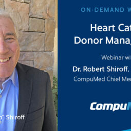
Scroll Top

How Serial Echos Can Improve Utilization: Case Studies

EXPERT WEBINAR SERIES

With Expert Cardiologist Dr. Robert Shiroff

FREE On-Demand Webinar

Dr. Robert Shiroff discusses the benefits of serial echos, when to do them, and how they can improve utilization. In some cases, when a heart has received an insult as a result of brain death, trauma or other factors, a series of echos can help determine if a heart is recovering, or if the damage is too much to transplant. Drawing from multiple case studies, Dr. Shiroff will show how tracking heart progress over time with a series of echos can be beneficial during donor management in determining a viable heart for transplant.

Who is This For?

Donor & Transplant Professionals

00:00:00:00 – Laura Carroll
Today, we’re honored to have again Dr. Robert Shiroff, CompuMed’s medical director, to present some information on how serial echos may help save more lives and increase the number of viable hearts for transplant. I think most of you on this already know Dr. Shiroff may have talked to him at 2:00 in the morning. When you’re managing a donor case.

00:00:20:19
And what you might not know is that according to CompuMed’s estimates, Dr. Shiroff has read the most donor echos ever than any other doctor, including all of our other reading doctors at CompuMed. And, you know, he’s used this experience in all his other vast experience on a daily basis to help us improve our echo reading processes and our heart cath protocols that some of you are familiar with and saw that webinar also.

00:00:50:21
And so today he’s going to provide some information on serial echoes. And just a quick note during the session. Please go ahead and submit questions and at the end, time permitting, Dr. Shiroff will address this. And without that, Dr. Shiroff, please go ahead and tell us more about serial echos.

00:01:07:14 – Dr. Shiroff
Thanks, Laura. And thanks for everyone for participating today and coming in, listening to my spiel. Basically, the question is why do we do serial echo? And the reason is that there’s an expectation that left ventricular systolic function will improve with time in a brain to an individual or even a DCD patient. The other side of the coin is why not to do serial echos?

00:01:35:22
Sounds simplistic, but if the initial echocardiogram shows the heart is transplantable and there’s no reason to repeat an echo. The other reasons not to do them. And this one is a hard one for a lot of folks, is that the resistance the hospitals perform timely serial echos. This gets down to the basic issue of technician time and for a dead patient in the hospital’s resistance to provide the care in a timely fashion.

00:02:03:03
Because we need echoes at six or 12 or 24 hour repeats, possibly a couple of times. And they wonder why we’re bothering to do this. The last the third one is cost considerations. And besides the cost of our services and the cost of doing an echo, the ICU utilization is a big one for hospitals in the system.

00:02:24:15
Obviously, you’re tying up an ICU bed and if you have not had the heart recovery or organ recovery for the patient, you continue to utilize that bed while you’re waiting. The other is the cost of the study in delaying other organ transplants and potentially losing them. And affecting the organ transplants as time goes on. And we know that hemodynamics can deteriorate and kidneys can be injured and may be lost for transplant.

00:02:52:04
Next one is legal considerations. Sounds ridiculous, but there are situations which where homicide is questioned or there’s questionable cause of death and the coroners or the district attorneys or other legal entities get involved in the cases and they don’t want to wait for 24 or 40 more hours. The last one is social issues. And this involves a family that has been had a blow, obviously, to them emotionally that their loved one is legally dead.

00:03:20:00
But sitting there and with a ventilator going. And the question is how long we have to wait till we can take care of dealing with our own grief. So this becomes an issue and a significant one for families. Let’s first talk about the heart function, what we’re talking about ejection fraction. When I did a dress rehearsal this couple of days ago, someone said, well, how do you define ejection fraction?

00:03:45:03
And I realized I hadn’t defined it. So I put the slide in and basically it’s the performance of the heart. And the heart contracts. A certain amount of blood is left in the heart. What is injected with each beat is called the ejection fraction is put as a percentage. It’s very simply the end diastolic volume is you take the end systolic volume and you subtract this man diastolic volume.

00:04:05:13
You put it over the and diastolic volume and you multiply by 100. Well, how do you measure that in echocardiography, which is the most common way it’s measured, because it’s so easy to do. There are at least three different techniques for measuring it. One is called Teicholz one is called Simpson. Teicholz is basically an ice pick view and a two dimensional echo.

00:04:26:10
And you’re measuring walls moving toward or away from each other and you get the measurements and you make certain assumptions physically about a cylinder revolution, which is more complicated than you need to know about. But from that information you calculate an ejection fraction. The next way is with Simpson, which is a volumetric measurement where they actually try and measure the volume of the ventricle by drawing and I diastolic picture in in various views of the four chamber and two chamber and short axis and even left parasternal view as well.

00:05:01:24
In that circumstance, another equation is used to determine what the ejection fraction is. The third one is in 3D echoes, which does it by computer and more mumbo jumbo than I care to go into it right now. In addition to that, there’s other ways to measure ejection fraction. It can be measured with heart catheterization when you shoot, when you do a ventriculogram and die is put in the left ventricle and you watch in RAO projection, particularly, the operator will draw a picture of the ventricle and diastole and and systole and the computer will then do an computer analysis and come up with an ejection fraction based again on a bunch of equations.

00:05:42:18
There’s also three more methods for measuring ejection fraction. You can do it through MRI, you can do it through CT and you can do it through nuclear. You ask why these all these measurements available and there’s so many different ones. Well, my experience in medicine has been when there’s a lot of different ways of doing something, it tells you that none of them is perfect.

00:06:01:00
And that’s the case. That’s why. And when you read an echocardiogram and when I read an echocardiogram, it has a box says visual ejection fraction. That’s an estimate of the experienced cardiologist view of the heart and how what the ejection fraction is. Most of the time we agree pretty much. But when you have segmental dysfunction and when you have other issues of the heart, you can disagree significantly with a measured ejection fraction.

00:06:24:24
So I’ll stop there about ejection fraction, because that’s more than I ever wanted to talk about it. Well, why the expectation for improvement of ejection fraction in a braindead individual? And that depends on the ideology of the ejection fraction of the decreased ejection fraction. And probably the biggest, most important part of this whole thing is downtime. So if someone is injured in a trauma, injured with a cardiac arrest, secondary to an overdose of cardiac arrest, secondary to hanging two other suicide attempts or a CNS bleed, it matters with what with etiology is in, as well as if you have a cardiomyopathy of Takotsubo which we’ll talk about in a minute.

00:07:10:20
Well, let’s talk about CNS bleeds for a minute. First is the blood pressure instability And those of you who’ve cared for patients with subarachnoid hemorrhages and bleeding from an aneurysm acutely kills people because they herniation they get marked hypertension with that blood pressures in the two fifties over one thirtys, one fourtys and if they’re going to survive which our patients have not obviously, but if they’re going to survive, blood pressure should be lowered very quickly, lowered very quickly.

00:07:40:02
LV Dysfunction occurs secondary to blood pressure rises and also secondary to myocardial injury that can occur with subepicardial ischemia. And there are newer humeral effects associated with intracranial bleeds that are complicated and not well understood, but can cause LV dysfunction, which can be transient. And we’ve seen a lots of recovery with the CNS bleeds.

00:08:01:15
And this is one of the areas which we like to highlight and think seriously about doing serial on. cardiac arrest is, as I mentioned earlier, the most important thing is downtime. And I discovered in trying to come up with slides and pictures to show you of the echocardiogram since taken serially by our people here at CompuMed and read serially by CompuMed cardiologists is that we have not the kind of data we should have.

00:08:29:19
We don’t have a database and we need to form that. I think it’s going to be up to the OPOs to think seriously about getting a standardized form where we can document what the cause of the persons of the patient’s brain death is or CD, but death is when it occurs how long they were down, if they had downtime, what the cause of the downtime was.

00:08:52:12
If you have that irrelevant and other issues which will then allow us to come up with some better handle on how to better handle this secondary to trauma, cardiac arrest is secondary to hypertension, hyperkalemia and shock, which can cause brain injury because without the decreased perfusion in the head and swelling and herniation, secondary to drug effects with drug overdoses, and you will go through some of the various drugs and the problems inherent in those particular entities. suicide attempts, so many times with respiratory depression, with opioids, with hangings, you have trauma to the trachea and you have respiratory arrest as well.

00:09:31:22
And that’s the primary cause of death. Cardiac depression occurs with respiratory depression and hypoxemia and Takotsubo cardiomyopathy, called so-called sad heart syndrome, is a segmental dysfunction which occurs in in depressed patients, which goes back to suicide attempts. And in that circumstance, we would see an abnormal ventricular function. And in the outpatient setting or inpatient setting in patients who have survived or gotten to the hospital without having a brain death or an arrest, with Takotsubo they will many times get better with conservative care.

00:10:08:00
And the question is, can we make them better in brain death individuals? And that’s yet to be determined. There’s some people that are doing some studies looking at that now, from my understanding and some trials being true, some throw at the wall and see what happens with the kind of events. Let’s talk about the drugs for a minute, if we may.

00:10:25:10
Opioid effects in the heart are many direct and indirect. The direct effect is infective Endocarditis occurs with the opioid, with intravenous drug use of any kind. So I just throw that in the opioid category. But that can be can be thrown into the methamphetamine category as well. But usually we see that on an echocardiogram. I’ll tell you that we recently had a case that I read that I, I reviewed and three other cardiologists reviewed where I did not see the endocarditis.

00:10:52:24
And the surgeon after heart recovery appropriately looked at the heart looking through the atrial appendage. He said they saw the left atrial whereas cut off from the from the donor and looking into the heart and looking at the mitral valve saw some more vegetation. We went back and looked the echoes and couldn’t see it and we could not see the small hole that was reported as well.

00:11:17:02
The interesting thing was this echo was the last echo was done more than 48 hours after the one that we had read is not having endocarditis that it occurred during that timeframe. I don’t know. Probably not. We probably just missed it because it wasn’t this test is not sensitive enough to pick up small lesions. That’s one of the reasons you have to keep checking these hearts when even after they are recovered.

00:11:38:07
The other thing it happened, direct effect of opioids is prolong QT interval. QT interval. It leads directly to the prolonged duty interval directly to this arrhythmias like Torsades de pointes, which is like in between ventricular tachycardia and ventricular fibrillation can lead to death. Decreased cardiac output can occur with direct effect of the drugs and bradycardia can occur.

00:12:02:17
And we’re talking about OxyContin. Heroin not fentanyl bradycardia occurs as well. The indirect effects are respiratory depression, which is what we deal with most of the time when patients don’t survive and cerebral depression. So opioids are bad drugs to have in this in the system. The question is, as they wash out of the system, if the patient is brain dead, what is the expectation that the negative effects on the cardiac status will be improved with time and it depends on the downtime to a large degree as well as the amount of drug involved.

00:12:36:10
Methamphetamines are bad actors all across the board. Their use is rising, is rising globally, resulting in significant morbidity and mortality driven by a poorly understood increase in multiple forms of cardiovascular disease. That’s a mouthful, but it’s true. It may provide cardiovascular disease through catecholamine toxicity. That is the increase in adrenaline and noradrenaline, and we’ll see that with cocaine as well.

00:13:02:04
And less effects through cardiac effects, through the direct effect on the cardiac system and the vascular tissue. The use of methamphetamine is associated with pulmonary hypertension, particularly in patients who have a specific genetic abnormality or genetic finding with polymorphism in the metanephrine catabolizing enzyme carboxylesterase number one, obviously we don’t know that one, and that’s a genetic test.

00:13:29:22
And we have to do right heart cath or look at the tricuspid regurgitation or tricuspid flow across the valve to estimate pulmonary pressures in the setting of methamphetamine. Next in this issue is symptoms of myocardial infarction with methamphetamine result from either acute cardiac vasospasm or enhanced atherosclerotic plaque formation. So chronic methamphetamine abusers get more heart disease, more coronary artery disease as well as vasospasm.

00:14:01:14
This is a an issue that leads us to leads a lot of times transplant centers after the decision to request heart recovery to proceed on and ask for a heart catheterization. And the coronary arteries are particularly interesting to those centers along with the PA pressures. The last thing is remodeling of cardiac tissue, where the current heart left ventricle dilates to a cardiomyopathy and may enhance susceptibility to cardiac rhythms.

00:14:30:06
We don’t usually have a problem with this if we get an adequate echo because a dilated cardiomyopathy is one that usually is not reversible, it’s takes a while to develop a cardiomyopathy that’s with dilatation, and therefore we don’t usually expect it to resolve in the timeframe required to heart, to recover the heart. The last bad actor I’m going to talk about is cocaine, and it’s a bad actor.

00:14:58:08
You can see a nice little good diagram here, although all roads lead to death, sympathetic output and catecholamine toxicity leads to increased heart rate, increased blood pressure, increase contractility, or improve ejection fraction. And that setting leads to ischemia of the myocardium because the heart rate is so excessive and the blood pressure is so high and the contractility is so high, the so-called double product blood pressure times heart rate determines to a large degree oxygen requirements is O2 requirements increase.

00:15:26:01
You get relative ischemia of the heart, which can lead with the setting of vasoconstriction and coronary spasm, two ischemia or infarction. And we obviously are already dealing with the death of the patient, possibly through this mechanism where possibly through a local anesthetic effect where they get rhythm disturbances with again QT prolongation QRS widening with ventricular rhythm disturbances associated with that which leads to LV dysfunction and to death via various mechanisms had already been defined here.

00:15:56:11
So cocaine’s a bad actor. This is why so many centers now we’re asking for that for heart catheterization to be performed on donors and on hearts and why we’re reading more those for you folks. Someone just slipped the question on the board in regard to my screen. This is a now I’m going to start looking at echocardiograms.

00:16:24:03
And we have three cases, and I wish I had better histories for you because I don’t I think this one was a gunshot to the head. But I’m not 100% positive and I won’t stand by it. This is an echocardiogram. Here we see the left ventricle. This is left ventricular contraction. This is the septum up here moving and should be moving in toward the arrow I’m showing you.

00:16:46:12
And the posterior wall should be moving in toward the middle and if you had an ice pick view, you would stop it. And you diagram it. Measure the systole and diastole and systole and diastole, and you do the equation that’s used for Teichholz. This is the left atrium here again, it’s anatomy. So you remember from last time at a time before where you have the mitral valve, but blood flows in from the left atrium to the left ventricle, then pumped out through the left ventricle, through the aortic valve, to the aorta, into the body.

00:17:18:05
This is a depressed, depressed ejection fraction. It was estimated 20 to 25%. We cheated over here to show you what these are, and that’s from based on a report performed by one of one of the other readers for CompuMed. This is another view is the four chamber to show you this heart is not contracting well here again it should be moving this toward toward the center of the chamber and you can see this moves a little bit, but not very well.

00:17:44:19
And one of the things it’s nice to have or always on an echocardiogram or an angiogram, is having an EKG. So you can see Systole and diastole really clearly see when the heart is coming in and when it’s not, when it’s relaxing. Is the mitral valve here left atrium? This is the right atrium, tricuspid valve and the right ventricle.

00:18:01:20
You don’t see the outer wall, the interior, while the right ventricle is called a for it’s almost a good four chamber. And you’ll notice these echos and they’re not all perfect. That’s the nature of the beast with echocardiography, particularly in patients who are ill in the ICU ventilated, and all the rest of the things that we were dealing with.

00:18:21:02
They’ve had trauma, they’ve chest tubes, they have subcutaneous emphysema. They have all kinds of issues that make echo, trans thoracic echo difficult and then challenging. This is a report from the first study that was done by Dr. Simpson. He called it 20 to 25%, which we showed you up here. The next series is that was done several hours later.

00:18:41:23
You can see a little better symptoms moving in a little more. And this is called 40 to 45%. I’ll show you the four chamber, this one as well. Again, I think it sees more vigorous contraction. And this one was called 40 to 45% by Dr. Simpson. Again, we try and have the same reader read them off if we can, if at this time, if time and circumstance allows, it’s good for continuity.

00:19:10:08
But if not, we’re all very, very close to reading and the experience level is really good. This is the third echo in the same patient. This one’s 50 to 55%. See how much more vigorous the septum is. And one of the things we did we learned from getting this together this time is we don’t have the technology available to us yet to be able to show you side by sides of the serial echoes of the same view.

00:19:33:15
Many times of use are identical where there’s close as we can get, but you can get thinking about ballpark that and see how much better that is. In the first echo that you saw, this is close to a four chamber view and again, 50 to 55 is probably reasonable for this one. And this one, there were three echos time.

00:19:51:22
There was a fourth one done in the middle. We didn’t get to read, but you can see the time frame here in our zero. When the first Echo was done, it was even got 25 to 30%, 20, 25%. Record is 20 is measured is 26. I’m sorry about that. I’m technology challenged, sometimes 42% for the second echo and 55% for the third echo.

00:20:18:15
And this was over 77 hours. And I read the third one next series begins with a little better echo, slower heart rate. And we have the EKG on this one. Same kind of view, the same parasternal long. This is close to four chambers. I could get on this one. And we this is called 45 to 50%. It was read by Dr. Simpson.

00:20:54:21
Again, see, this is improved here. This is this is more 55 to 60 is contracting much more vigorously. This is clearly a normally contracting heart, not exactly the same views, but close this. We only have two for our series, but you can see the difference in ejection fraction went from 42 to 59% over 28 hours. So this one improved dramatically over a shorter period of time.

00:21:29:15
And the other the first one. third study, this one we call 40 to 45%. Same views, a little faster heart rate. When we read them, we slowly we sometimes I slowed the heart rate down and get a better look at the wall motion but the it’s the technician can stop it when they want to. We have to stop it when we can.

00:22:05:05
When we’re reading these things. This one was read by my myself and called it 40 to 45. Second Echo, a more vigorous lot, a lot of artifact here. Looks like the ventilator is set at high frequency ventilation or or at least a faster rate of ventilatory rate. And you’re seeing the lung tissue coming the way of the transducer.

00:22:30:03
But I think you can appreciate that it’s improved the ejection fraction. I called it 50 to 55% or I think I was me stand for chamber. Again, still tachycardic, but you can see how much more aggressive that heart is in beating it compared the first one and the second case second and a third one in the same patient a little better again, again, difficulty with the ventilator.

00:22:59:22
If we get one or two beats we can look at, we can make a judgment and the technician stops it when it’s best for them, The measured to their measurements. And we said 55 to 60 on this one. Again, here is three time intervals up to 51 hours on this one. So there’s a long interval between the 24, one hour one and 51 hour one, but not that much improvement.

00:23:28:00
We drew the line, the yellow line at 50 per 50% ejection fraction. Some people call that normal. Some centers require 55% for a transplant. A lot of centers will look at the heart at 50% and with the hope and expectation that they can get it, you may get some more better, some better. We’ll stop here for your questions.

00:23:47:05
And there were some questions submitted earlier. And at your request, the questions were put to us and I’m going to try and answer them. first one is how long should you give a heart to recover? It’s a great question, and I didn’t do that. That wasn’t me. How long should you give a heart to recover?

00:24:07:11
Well, the ideal world, you’d go to Max and you do it forever until you got a heart you could transplant if it was possible. Obviously, that’s not practical. And I think the the limitation ones are related to those. Why not do serial echo type of things? It’s other organs at risk. It’s the cost in all the ways I listed before of waiting to do it, to do a recovery of the heart.

00:24:36:19
If it’s doable. I think we need more information is to know what the optimal time is and their given circumstance is. It may be different for different for different circumstances, and that’s my guess. Frankly, it’s going to be the case. So it’s a great question. I don’t think we have the answers. I think if you have a heart with it, that was the downtime was reasonable.

00:24:58:17
By that I mean 30 minutes or less. And you have a stable rhythm and you have some you got an early echo that showed 30 or 40% ejection fraction and you have the wherewithal to get a second echo at 12 hours or 24 hours. I would strongly suggest that that’s going to recover at that time. It may give you a trend that it’s showing signs of recovery, which will then encourage us to urge your supervisors, to wait another 12 hours and repeated again, because as we always mentioned, heart to heart say this is a life saved.

00:25:31:06
What can I do as a donor coordinator to help the heart recover of anything? Well, I think you have a very. Why is it do that to me? I think that the answer to that is that you try and keep the donor as hemo dynamically stable as possible, optimize fluids, decrease the catecholamines if you can, by reducing pressors if possible, make things as good as possible for this person as if they were alive from a hemodynamic point of view.

00:26:03:22
So you want good filling pressures, you want adequate volume load, you want adequate maintenance of blood pressure and respiratory needs, you want them well oxygenated and then you say a prayer. That’s basically where you’re at. What pressures are best for heart while trying to recover or certainly with whatever pressure will cause or adequate. I am sorry news. Keep skipping ahead on me and I don’t mean to be doing that.

00:26:32:11
What pressures are best for the heart while trying to recover? I think that clearly pressures that allow organ perfusion, if you’re making good urine, you’re perfused in the kidneys. That pressure’s adequate. If someone is known to be hypertensive and in life and their blood pressure is running 90, that may not be an adequate blood pressure. If their blood pressure, if if, however, at 90 they’re adequately profusing their kidneys, then I would then lower the blood pressure is the longer the better than it is for the heart in terms of the work required.

00:27:02:13
So the lower the after load and the more optimal the preload for the heart that is the venous return, the better the heart’s going to do in terms of chance giving a chance for it to recover. And as I say, keeping adequate oxygenation is critical as well. This is one that I put in. Not expecting too many questions.

00:27:24:11
Should we consider a multicenter study or information gathering to determine which hearts with low ejection fraction will improve? In which with time, with time and intervention? I think that there’s nothing else my doing this, getting this talk together and got me and started educating me that we don’t have the data. We I knew that we didn’t have data about how to predict when hearts are going to recover and what would make them recover.

00:27:52:16
But I know we don’t the data even before that to start knowing what questions to ask, we don’t know. We don’t have the database available yet, which I think we need to do. And I’m prepared to like we did with the cath information sheet where we ask cardiologists to minimum to minimum things required for heart.

00:28:12:24
Cath I’m prepared to draft a document with it with some help from coordinators or other physicians in the up in the transplant community of what data we need about potential donor hearts and and what’s happened to the patient. Because I think our chance of bringing back more hearts and having more donor heart recovery is going to be dependent upon more information about what happened to that heart and individual and what was done to them.

00:28:42:21
So in other words, right now t four is used across the board almost for if for donor heart rate time of before transplant. A lot of people are using dobutamine as a way of trying to assess whether it’s going to recover by why we say is whipping the heart because it’s an inotrope and chronotrope. The other thing is for Takotsubo.

00:29:01:16
Some people are using estrogen now in an attempt to try and prove myocardial function with estrogen given in high doses for a couple of the to one or two days. I don’t know if any of these are going to work, but I think we need more cooperation in the community to gather data, at least get some preliminary trial going.

00:29:20:00
When we look at that historical data, don’t change what you’re doing, won’t make anybody be forced into a protocol, but accumulate data on what various centers are doing and see what works and what doesn’t work. Everybody believes what they’re doing is making a difference. Maybe there’s somebody out there doing something that’s making more of a difference, and I would encourage that type of approach at this point in time.

00:29:40:18
And I’ll leave two more questions. Any more questions, Laura.

00:29:43:06 – Laura Carroll
That we do. So I’ll just go ahead and read this. The first one is submitted by Daniel Stouter. He says, Could you please touch on the challenges presented by echocardiograms with sub optimal image quality? How do you typically I know there’s a more how do you typically address that you utilize an injectable enhancement agent, i.e. lipid coated echo genic, micro bubble based products, or do you opt for a different modality entirely?

00:30:09:23 – Dr. Shiroff
Well, I think if it’s just a question of what ejection fraction is, if adequate could look at the valves because obviously with drug with drugs out there, endocarditis is an issue and you want to make sure you’re not dealing with congenitally or some other unusual valvular abnormality. If you have adequate look at the valves on a trans thoracic echo.

00:30:29:10
Well, the only thing you’re missing is wall motion. And you can’t determine wall motion because it’s such a poor quality study. In contrast, studies echo contrast is of significant value. And even without you, even with valves not being perfect, if you have a 25 year old, if you get a one glimpse of the of a mitral valve in the tricuspid valve, in the aortic valve, you probably kind of say it’s probably okay.

00:30:52:03
But I think that and that circumstance where there’s really a poor enough quality echo to another modality, transesophageal echo should give you high quality imaging of wall motion as well as valvular anatomy and looking for leaks or stenosis So it’s it is a judgment call. And we will recommend to you which we think should be done in the echo report and were verbally.

00:31:18:22
And so you know what their thoughts are.

00:31:24:09 – Laura Carroll
Our next question is from Alisha Jackson. And she asked us how often or are OPOs using definity for difficult studies? If so, if so, is there available training for that? Are there benefits outside of visualization difficult study?

00:31:41:15 – Dr. Shiroff
Well, that’s the same basic almost the same question because they echo contrasting and that’s fine. Definity is one of them. There’s there’s there’s several others off the con and there’s several others out there. It depends on what the hospital that you’re working in is using because usually have a contract with one company because of volume. And it certainly does help you in terms of defining wall motion and also can help define, well, thickness because you see a negative image in and that’s that’s usually the septum and posterior wall if you can get an adequate view of it.

00:32:10:06 – 00:32:33:12
Dr. Shiroff
So yeah, it doesn’t help where you have subcutaneous air. Nothing, nothing helps in that circumstance. If it’s trauma patient or event long term, long term ventilator patient, those patients, you can’t yeah, you need to be able to get sound waves to get to the heart and bounce back in a somewhat clean fashion. So contrast material does help. And sometimes and sometimes it doesn’t.

00:32:35:07 – 00:32:54:16
Laura Carroll
We have another question here, and a lot of them, I think are this is some of these similar and as I always say, full disclosure, I’m not clinical. I’m just reading these to were any interventions besides time utilized on the case study shown points on T4 inotropes?

00:32:55:03
Yeah. I’m not sure.

00:32:55:17 – Laura Carroll
Which patients.

00:32:57:17 – Dr. Shiroff
And clearly I don’t know.

00:33:00:12 – Laura Carroll
Yeah, that’s the challenge, right? That’s why you pick up the phone and call sometime.

00:33:04:22 – Dr. Shiroff
And it’s exactly why I get on the phone, talked to coordinators is the what? You know, the first question I asked, what happened to them and when did it happen? How long ago and how much was down. One was a downtime because if you if you want an opinion about what and what was the cause of it and whether it was one of the drugs that we mentioned or something else, you know, and I think that and how many days ago it occurred when five first Echo was done, five days after cardiac arrest and the ventricles, terrible is probably not going to get better no matter what you do.

00:33:33:24
If, however, it’s 12 hours after cardiac arrest because of a of a hanging or a drug overdose and the downtime is unknown. It was 30 minutes or somebody said it was an hour. It’s worth, if you can, to try some of these modalities and we don’t have the adequate histories. And I was very disappointed in myself in not coming up with a way of doing this.

00:33:56:08
But this is focused that focused me in this whole area of getting more information and CompuMed’s going to try and push the window of getting more information from the centers, if we can, so we get them a data database, but be great if it could be shared between OPOs rather than and how and how we do that. I’ll leave it up to the higher power as.

00:34:15:24 – Laura Carroll
Well and sharing information on the front end. And then also on, you know, was was it transparent? So that we can start to track that differently. So it’s both advice.

00:34:25:21 – Dr. Shiroff
That is exactly right. But the bottom line is we don’t know what these were trans. I know one of them was the first one I did was definitely transplant because I was told that by the coordinator. But beyond that, I would I do not know whether there’s other ones were transplanted. I can assume. But, you know, assume does it makes a as I said, you and me.

00:34:44:03
And so you just.

00:34:47:14 – Laura Carroll
And I will jump in on that case. You mentioned earlier that we read and we actually talked with the medical director at that OPO And we actually love follow up questions for anybody that’s on this and doing a post Q&A, if you will. We have all of our other reading Doctors read it. We’ll have 4 to 5 of the cardiologists look at it, and we learned quite a bit from that.

00:35:11:17
They went back to the transplant hospital. So, you know, we love to do QA after the fact, if anybody wants to submit this to us. So I’m just throwing it out there. More work for everybody. But we love to do it.

00:35:21:21 – Dr. Shiroff
Absolutely.

00:35:22:23 – Laura Carroll
Yeah, we learned. So another question and I think these have come up in other webinars we’ve done. There are many differing opinions on which pressors should be weaned first at the donors on multiple pressors. What are your thoughts?

00:35:38:07 – Dr. Shiroff
Yes, there there it is a matter of this, a matter of style as much as anything else. The medications that increase O2 consumption of the heart, but that is the ones that caused more tachycardia are ones that I would prefer to get off first. The ones that ease the work on the heart are the ones I would get off last that you have to maintain blood pressure, mean, mean arterial blood pressure and you have to maintain perfusion in the organs.

00:36:06:11
So you do what you have to do. I mean, when I was practicing on live people in the ICU for years and years and years, the final arbiter in terms of keeping people alive so they could talk to their visit with family members before they died was a drip of epinephrine and calcium. And people would be are appalled by that combination because it’s maximal oxygen consumption.

00:36:30:22
But it kept them that they allowed them to perfused their head enough to be able to say goodbye to loved ones. And I found that was useful in that setting because this patient was going to die regardless of what we did. Yeah, keeping a dose of dobutamine down to non tachycardic doses is better than having it at higher dose and dopamine the same thing it we try and avoid levophed if it you’re trying but if you put in certain circumstances, if it works well you’re trying to use milrinone if you can.

00:37:03:00
It is complex and depends on the individual situation and has to be evaluated at the time and you need a sophisticated intensivist to help you with that. And I want a data on a moment to moment basis.

00:37:16:21 – Laura Carroll
Well you actually just pretty answer the next question, but I want to bring it up again. So Michael Williams asked, you mentioned domutamine t4 estrogen. Is there utility for no or no? No, I know how to say it because you just said it. So yeah, you can expand on that a little bit more.

00:37:33:20 – Dr. Shiroff
Well, there is in certain circumstances, but again, we don’t have all that typically in patients where we use drugs like milrinone we have hemodynamic monitoring devices in their way. There are a lot of patients that you’re trying to get if compensated from decompensated situations in the setting of a potentially recovered heart or the transplanted heart. The you don’t have that.

00:37:55:24
So you’re working in the dark. So it’s a lot to use as complex drugs that rely on knowing what the filling pressures are when you when you infuse them. So it becomes problematic, you know, so I think that’s one of the reasons they’re not used that much, because it is a difficult problem to try and pick the right dose.

00:38:14:10
It’s not a like a shopping cart and you just put in 2.5 miles per kilo per minute or whatever of drug you’re using. You have to know what you’re doing and how you’re doing it and why you’re doing it.

00:38:30:12 – Laura Carroll
Right now there are no more questions. So we purposely kept the this presentation part of this at half an hour because we know, especially in the OPO transplant world, you guys are super busy. So we’re trying to keep this a little bit more staccato than an hour long. If you have more questions, go ahead and contact us. There’s a few contacts on this last slide.

00:38:52:19
And I think at the end of this, you can let us know if you want to reach out. I peruse the names and I see a lot of names on this that are current customers. And so you know where to reach us. For others, the information’s on here. We’d love to share more with you. This will be sent back out to everybody that registered for a rewatch of it.

00:39:13:16
Feel free to share it with your your colleagues. And actually, if you go to our website, you can see all our part, some of our prior presentations on there too. We had Dr. Simpson talking about pediatric cardiology and the donor management of that. We had a doctor Angel talking about pulmonology. A couple of times Dr. Shroff has done he did one on heart Cas and in December, late December, early January, we’re going to be doing some pathology and then we’ll have a whole calendar again for 2023, if you can believe that.

00:39:45:03
It’s already that time of the year. We really appreciate everybody being on the call and for your time. And again, just reach out to us and let us know how we can help you save more lives also. Thank you.

Robert "Bob" Shiroff, MD, FACC

CompuMed Chief Medical Director
Clinical Cardiologist

Dr. Shiroff is a board certified cardiologist with a fellowship in clinical pharmacology as well as an experienced medical expert witness.

CompuMed is honored to have Dr. Shiroff on our team as he serves the donor and transplant community. His in-depth knowledge and expertise helps assess, manage, and improve the recovery of donor hearts, resulting in an increase in the number transplanted into needy recipients. Knowing that each recovered heart has the opportunity to save a life is humbling and extremely rewarding. We are grateful for his passion to share his knowledge and experience with the OPO and Transplant community.

Privacy Preferences
When you visit our website, it may store information through your browser from specific services, usually in form of cookies. Here you can change your privacy preferences. Please note that blocking some types of cookies may impact your experience on our website and the services we offer.