What is freezing artifact, and how can we identify it?
To minimize freezing artifact, we must first understand what it is and why it’s detrimental to an organ read. Freezing artifact is caused by ice crystals that disturb the tissue sample during the freezing process. Here’s an overview of four types of freezing artifact and how you can identify them.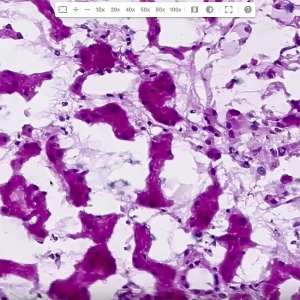
Compression Artifact
- Bubbling artifact. As mentioned above, one type of freezing artifact is described as looking like Swiss cheese, with bubble-like spaces within the layers of the tissue.
- Compression artifact. One of the most frequent types of freezing artifact, compression artifact is characterized by a squeezed or squished appearance of the tissue’s tubules.
- Chromatin changes. Chromatin changes involve the alteration of the appearance of nuclear material, often making the chromatin (the substance within a cell nucleus) appear clumped or fragmented.
- Nuclear holes. Nuclear holes are observed as empty spaces within the nuclei, making them look hollow or incomplete.
How do I minimize freezing artifact?
The formation of ice crystals is the primary cause of freezing artifact. You can help prevent them by ensuring a rapid freezing process and using best practices for creating frozen section slides, such as: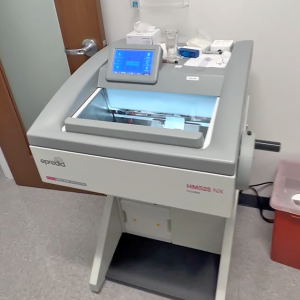
Cryostat
- Ensuring cryostat temperature is set to -24 degrees.
- Reaching the ideal cutting temperature, which is achieved when the block is between -17 and -18 degrees. Two ways to ensure proper cutting temperature include:
- Utilizing a temperature gun.
- Checking the color of the OCT (Optimal Cutting Temperature) compound, which should be a solid white. If the OCT media is gray in color, this is a reliable visual indicator that it hasn’t reached the ideal cutting temperature.
- Slicing tissue samples to 5 microns, the gold standard in thickness.
- Performing preventative maintenance on equipment and hardware at least every six months.
3 Tips for Troubleshooting
Experience and proper training can also help you become proficient at troubleshooting issues that lead to freezing artifact. Fernando offers three tips to help optimize your slide preparation techniques to minimize its occurrence.- Learn the differences between core biopsies and wedge biopsies.
Knowing what kind of tissue you’re looking at will help minimize freezing artifact. “Different organs have different water content, so if you are familiar with the water content of the tissue you’re dealing with, you can say whether the water content is going to be more than normal,” says Fernando.
- Pre-chill your OCT.
Pre-chilling your OCT (optimal cutting temperature) compound can also help minimize freezing artifact. Fernando recommends keeping OCT in a controlled environment, such as a nearby refrigerator set at 4 to 6 degrees. He says a refrigerator is preferable to the cryostat, as you may not know exactly when the tissue sample will arrive.
- Place well bars, chucks and heat extractors at the bottom of the cryostat.
Additionally, well bars, chucks and heat extractors should be placed at the bottom of the cryostat to stay at the lowest temperature possible. This is significant because the tissue sample will need to be frozen rapidly, and the longer the process takes, the more freezing artifact will appear in the section.
Focusing on these tips will help rapidly freeze the sample and minimize the amount of freezing artifact. As Fernando says, “You’ll get to the point that you’ll know when cutting a section, okay, this isn’t going to end well. You’ll start putting the dots together.” “It’s not just one reason or variable,” he adds. “Minimizing freezing artifact is something that comes with practice and observation.”