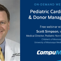
For infants, children and adolescents on the transplant waiting list, time is a significant challenge. Currently, about 25% of infants will run out of time before receiving a donor heart. The situation is only slightly more optimistic for older children and adolescents, with waitlist mortality rates around 15%.
As remote diagnostics partners to OPOs nationwide, our CompuMed sub-specialists understand the impact these statistics have on organ recovery teams. Managing pediatric donors presents unique challenges, but by implementing effective imaging and donor management strategies, we can make a meaningful difference together.
“One of the biggest blessings and amazing things about being involved with healthcare is that we are there for families in some of the most trying times in their lives,” says pediatric cardiology imaging specialist Dr. Scott Simpson. “OPOs definitely play a crucial role with that in the transplant realm — so how do we work together to get the waitlist and mortality rates down?”
In response to this critical question, Dr. Simpson shares nine key imaging and donor management recommendations to help optimize pediatric donor heart assessments and ultimately save more lives:
1. Normalize Vital Signs
Maintaining stable vital signs is crucial for an accurate assessment of ventricular performance that can lead to organ acceptance.
“I’ve had echocardiograms where the heart rate is 180 in a 3-year-old,” says Dr. Simpson. “That’s like trying to interpret a hummingbird heart — it’s beating so fast.”
Adjusting medication to stabilize heart rate and blood pressure can help ensure more reliable heart evaluations.
2. Reduce Pressor Use
Try to reduce reliance on vasopressors that can skew potential donor heart assessments.
“If I give a glowing report that the ventricular function has an ejection fraction (EF) of 70%, but the potential donor is on significant doses of vasopressin, epinephrine and other vasopressors, transplant teams can understandably question the true function of the heart,” explains Dr. Simpson.
Where possible, reducing pressor support gives a more accurate picture of a potential donor’s suitability for transplant.
3. Ensure Height/Weight are Provided
It’s essential that you include a patient’s height and weight. This data is used to generate body surface area for Z-scores, which help adjust heart measurements for pediatric patients.
4. Obtain a High-Quality Echocardiogram
A clear, detailed echocardiogram is essential to properly evaluating a potential pediatric donor’s heart.
“Quality in is quality out,” emphasizes Dr. Simpson.
5. Remove Unnecessary Devices on the Chest
One way to help obtain a high-quality echocardiogram is to remove all unnecessary devices on the patient’s chest, such as EKG stickers. This will help prevent obstructed imaging.
6. Utilize a Pediatric-Trained Sonographer/Fellow
Another way you can help ensure a high-quality echocardiogram is to utilize a pediatric-trained sonographer for the study, as they are more familiar with subtle things that will be emphasized in pediatric cardiology compared to adult cardiology.
7. Obtain a Complete Study (Usually >60 Images)
It’s also important to ensure a complete study is obtained for a full 360-degree view.
“In pediatrics, we are used to looking at the heart from many different angles,” says Dr. Simpson. “I’m expecting 60 images or more, sometimes even 120. Whatever you can do to ensure it’s a complete study – you don’t want the sonographer to rush it.”
8. Use Echocardiographic Contrast Agents When Needed
While not commonly used in pediatrics, contrast agents can significantly improve imaging in older teenagers or donors in early adulthood.
“If there’s chest wall trauma or the body habitus is unfavorable and it’s difficult to see the images, the use of echocardiographic contrast agents can be helpful to highlight the borders and makes getting the images much easier,” says Dr. Simpson.
9. Consider Transesophageal Echocardiography (TEE) in Certain Cases
In cases where chest wall trauma or other factors make standard imaging difficult, a TEE may be the best way forward.
“We would insert an echocardiographic probe down through the esophagus, which travels right behind the heart and eliminates obstructed viewing related to the lungs or bones,” says Dr. Simpson. “It has its own limitations but would be an excellent way to assess ventricular performance. I would suggest considering that in an appropriate setting.”
Optimizing Pediatric Donor Heart Assessments Saves Lives
Implementing these recommendations for pediatric cardiology imaging and donor management can help improve the accuracy of heart evaluations and allow transplant surgeons to make more confident decisions – such as saying “yes” to an organ that might otherwise have been declined.
Additionally, these recommendations can help honor the selfless gift of pediatric donor heroes. After all, the more hearts that are saved, the more families will receive hope and healing through donation and transplantation.
Explore More on Echocardiograms
If you found this information helpful, check out these other insightful case studies and tips on how serial echocardiograms can impact transplant outcomes:
- How Serial Echoes Help Capture Every Transplantable Organ: A Case Study by CompuMed Partner Live On Nebraska
- How Serial Echocardiograms Can Help Increase the Pool of Viable Hearts for Transplant
Learn how CompuMed supports OPOs throughout the donor management process. From the real-time, secure transfer of DICOM studies and organ recovery coordinator echocardiogram training to the 24/7/365 availability of interpretations and timely reports with hyperlinked data, CompuMed offers a variety of services and tools to enhance the donor management and allocation process. Connect with us today to learn more.


